Rob Gerritsen, specialist VIM (dipl KNMvD)
![]()

Together with John van Gulik, application specialist at Canon Medical Systems Europe (previously Toshiba), our team worked to develop an innovative contrast injection technique, which may be of great value to pets and vets (whether working in research or practice) worldwide. An abstract and poster with some first results were presented during a ct-user meeting in Ghent, Belgium (December 9-10, 2016).
Multiphasic contrast-enhanced CT is routinely used in human medicine to visualize hyper- and hypovascular tumours, hemangiomas and/or urinary tract disease. With this method a bolus of contrast is given to the patient and multiple scans are performed at set time points. The table will pass the gantry several times. Each scan will register and show the specific characteristics per respective hemodynamic phase (arterial, portal venous, delayed). Together, these scans provide a more reliable and relatively complete overview of what to expect with regard to a patient. A benefit which, for reasons explained below, is very relevant to oncology patients.
Veterinary medicine, with the exception perhaps of university clinics, did not exactly embrace the triphasic technique. Reviewing available literature, only an incidental study is found (f.i. Kutara et al. 2014, assessing the sensitivity and specificity of the technique in hepatic masses). The question is: 'Why?'.
The answer is rather simpel: multiphased contrast CT is challenging in animals, due to long scan times and necessary general anesthesia and breath-holds, especially in small animals. From a veterinary point of view the technique is complex. As a referral-only specialist clinic, we set out, together with Toshiba to develop and optimize an innovative 'split-bolus single-pass' contrast technique, which is used in human medicine to combine multiple phases in one single scan. The table will only pass the gantry once, after a native scan has been made; the diverse hemodynamic phases are captured in one single image.
 | 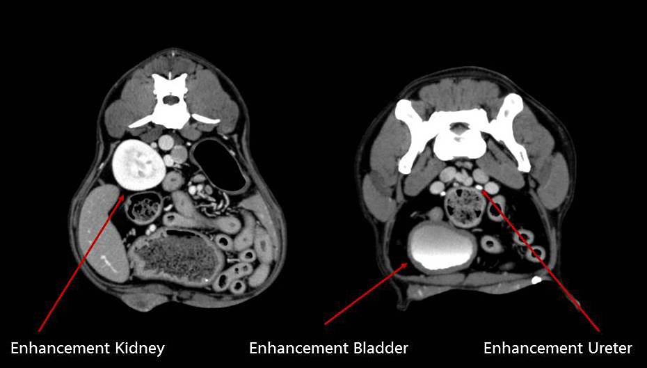 |
Photo 1 and 2: Axial split-bolus single-pass contrast enhanced images of a dog, 28 kgs | |
In veterinary practice untill now, usually only one phase is scanned, dependant upon the region of interest and the differential diagnosis. It is for that reason highly probable that essential diagnostic information is lost. Different tumours and their metastasis will show in various phases: some will be enhanced during the arterial phase, others during the portal venous or equilibrium phase. Every tumour, the primary and the metastasised, will be fed by an artery. Patterns of contrast enhancement will differ per tumour type, dependant upon the size of this artery, dependant upon the speed and level of blood supply, dependant upon the portal venous discharge and the enhancement of the tissue surrounding the primary tumour or metastasis.
In human oncology, reviewing all three phases is standard. Multiphased CT, together with clinical observations and pathology, provides essentail information necessary to determine type and stage of the tumour. Many studies into multiphased CT, have focussed on liver tissue. It was often the primary region for evaluation, due to the fact that it is the most important organ to host 'metastasis' due to abdominal tumours. Nevertheless, spleen, pancreas, stomach and colon may also be evaluated reliably and precise.
A pilot study was done. Using our Canon Aquilion CT scanner, we performed abdominal split-bolus 16 slice detector CT scans in a total of five privately owned healthy animals: four dogs and one cat.
By intravenous administration of two or three contrast boluses at specific intervals prior to the scan, we managed to register the arterial phase (HAP/AP), the portal venous phase (PVP) and the delayed phase (DP) in a single pass. For contrast medium, iohexol 300 mg/ml was used.
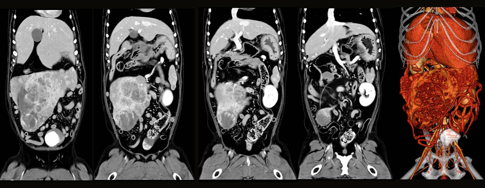 |
| Image 3: Rakker, Bernese Mountain dog, 38 kgs, split-bolus single-pass contrast enhancement. |
The images above, represent 'Rakker', an 11 year old Bernese Mountain dog, suffering from a spleen tumour (pathology after surgery indicating 'hemangiosarcoma'). We decided to use the split-bolustechnique. Above, separate views showing different stages of contrast enhancement of the tumour, with no indication for liver metastases. Rakker turned out to be one lucky dog. He was sent back to the refering vet for surgery and is still doing well.
We've invited practitioners in our area to help us refine and optimize our preliminary protocol by referring patients in need of abdominal and/or thoracic CT-examination to De Kompaan. We will be pursuing publication in a peer-reviewed journal.
Lees al onze referenties